|
|
Post by Vortex | Head Admin on Jul 12, 2015 22:37:40 GMT -5
Hey guys, this is a post about the types of brain imaging used today. Types of brain imaging This is an exact replica of this webpage which was written by Michael Demitri, M.D. I provide full citation below. 1 Brain imaging techniques allow doctors and researchers to view activity or problems within the human brain, without invasive neurosurgery. There are a number of accepted, safe imaging techniques in use today in research facilities and hospitals throughout the world. fMRI Functional magnetic resonance imaging, or fMRI, is a technique for measuring brain activity. It works by detecting the changes in blood oxygenation and flow that occur in response to neural activity – when a brain area is more active it consumes more oxygen and to meet this increased demand blood flow increases to the active area. fMRI can be used to produce activation maps showing which parts of the brain are involved in a particular mental process. CT Computed tomography (CT) scanning builds up a picture of the brain based on the differential absorption of X-rays. During a CT scan the subject lies on a table that slides in and out of a hollow, cylindrical apparatus. An x-ray source rides on a ring around the inside of the tube, with its beam aimed at the subjects head. After passing through the head, the beam is sampled by one of the many detectors that line the machine’s circumference. Images made using x-rays depend on the absorption of the beam by the tissue it passes through. Bone and hard tissue absorb x-rays well, air and water absorb very little and soft tissue is somewhere in between. Thus, CT scans reveal the gross features of the brain but do not resolve its structure well. PET Positron Emission Tomography (PET) uses trace amounts of short-lived radioactive material to map functional processes in the brain. When the material undergoes radioactive decay a positron is emitted, which can be picked up be the detector. Areas of high radioactivity are associated with brain activity. EEG Electroencephalography (EEG) is the measurement of the electrical activity of the brain by recording from electrodes placed on the scalp. The resulting traces are known as an electroencephalogram (EEG) and represent an electrical signal from a large number of neurons. EEGs are frequently used in experimentation because the process is non-invasive to the research subject. The EEG is capable of detecting changes in electrical activity in the brain on a millisecond-level. It is one of the few techniques available that has such high temporal resolution. A popular EEG device that is directed towards the average consumer is the Epoc by Emotiv Inc. Also, Mikhail Kabakov, has been researching, "Using EPOC to help create full-dive virtual reality technology, Full-dive VR, immersive VR, avatar control." You can find the thread here. MEG Magnetoencephalography (MEG) is an imaging technique used to measure the magnetic fields produced by electrical activity in the brain via extremely sensitive devices known as SQUIDs. These measurements are commonly used in both research and clinical settings. There are many uses for the MEG, including assisting surgeons in localizing a pathology, assisting researchers in determining the function of various parts of the brain, neurofeedback, and others. NIRS Near infrared spectroscopy is an optical technique for measuring blood oxygenation in the brain. It works by shining light in the near infrared part of the spectrum (700-900nm) through the skull and detecting how much the remerging light is attenuated. How much the light is attenuated depends on blood oxygenation and thus NIRS can provide an indirect measure of brain activity. Sources 1.) Demitri, Michael. "Types of Brain Imaging Techniques." Psych Central. Psych Central, 30 Jan. 2013. Web. Personal CommentsI am currently contacting some experts in this field for clarification on which would be the best for what we are trying to accomplish. Currently, the best way looks like to use a combination of EEG/MEG and MRI/fMRI. You can read about it here. I will update this post when I have a detailed report. Please feel free to add on to this post, we want to avoid duplicate threads. |
|
|
|
Post by yawegrimm on Jul 13, 2015 16:09:03 GMT -5
Hi, i'm a French student in neuropsychology and i've a very bad English but i'm going to try to explain here what i know.
Firstly, you have to know that all theese types of brain imaging are not very promising in our field of interrest.
fMRI, and NIRS and PET SCAN track an Hemodynamic activity, (blood activity in the brain), but there is a time lag between the moment a brain zone is activated and the moment there is a blood activity. So, if you think about your left arm, the fMRI will detect a blood activity something like 3/4 seconds after in the involved brain area. And this is not somthing technology improvement can resolve, it's juste that the blood activity is too slow to be a good indicator. So we can consider thant theese techniques have a good spacial resolution (because they can track activity almost evrywhere in the brain with a good precision) but a very bad time resolution.
For the EEG and the MEG, they've a very good time resolution (something about milisecond) because they track an electric or magnetic activity.
But theese activities are very tiny and we just track a global noise of the brain activity, and it cant allow us a good spacial precision. There is some paradigms like "P300" and "Motor imagery" whiwh allow us to control some very basics games, but these technology have no really a potential of improvement (eeg exist since something like 50 ies and have not relly being improved since). So, here, a good time resolution but a very bad spacial resolution.
There is some interresting techniques but, it's pretty invasive and consist to put some electrodes directly in the brain,or, at least "on" the brain (between the cranial bone and the brain like electrocorticography), and you guess there is a lot of ethical fences and the world is far to be ready for that.
Sorry for the very bad english, i hope you will understand at least a little bit. But globally, already known techniques are very weak and not very promising for game control.
|
|
|
|
Post by Vortex | Head Admin on Jul 13, 2015 18:23:10 GMT -5
Hey yawegrimm, thank you for your response, it is greatly appreciated. You are correct, none of the brain imaging methods I mentioned are what were are looking for. We need a good temporal resolution and a good spacial resolution to achieve a BCI like the nervegear. A while ago this made me wonder if a combination of brain inmaging techniques could be used for research done now. According to what I know there is nothing preventing multiple imaging techniques to be used in parallel(fMRI, EEG, NIRS and CT). We can combine the data and create a higher temporal & spatial resolution. I've contacted a few experts in the field to ask them about this and I'm currently waiting a response. This paper describes what I'm talking about. However I still contacted experts for their knowledge and advice. |
|
|
|
Post by Suu on Jul 13, 2015 21:44:12 GMT -5
Hey yawegrimm, thank you for your response, it is greatly appreciated. You are correct, none of the brain imaging methods I mentioned are what were are looking for. We need a good temporal resolution and a good spacial resolution to achieve a BCI like the nervegear. A while ago this made me wonder if a combination of brain inmaging techniques could be used for research done now. According to what I know there is nothing preventing multiple imaging techniques to be used in parallel(fMRI, EEG, NIRS and CT). We can combine the data and create a higher temporal & spatial resolution. I've contacted a few experts in the field to ask them about this and I'm currently waiting a response. This paper describes what I'm talking about. However I still contacted experts for their knowledge and advice. Hmmm It is nice to see some knowledgable people for once. EEG is beneficial in that it can detect electrical signals very fast and relatively easily, but as you know, it has an extremely low spatial resolution and is readily affected by background noise. So I dislike how it may be used for VR. I would rather be more interested in the developement of different contrasts for fMRI, for example, calcium ions have been proposed. |
|
|
|
Post by Vortex | Head Admin on Jul 13, 2015 23:15:48 GMT -5
Hey yawegrimm, thank you for your response, it is greatly appreciated. You are correct, none of the brain imaging methods I mentioned are what were are looking for. We need a good temporal resolution and a good spacial resolution to achieve a BCI like the nervegear. A while ago this made me wonder if a combination of brain inmaging techniques could be used for research done now. According to what I know there is nothing preventing multiple imaging techniques to be used in parallel(fMRI, EEG, NIRS and CT). We can combine the data and create a higher temporal & spatial resolution. I've contacted a few experts in the field to ask them about this and I'm currently waiting a response. This paper describes what I'm talking about. However I still contacted experts for their knowledge and advice. Hmmm It is nice to see some knowledgable people for once. EEG is beneficial in that it can detect electrical signals very fast and relatively easily, but as you know, it has an extremely low spatial resolution and is readily affected by background noise. So I dislike how it may be used for VR. I would rather be more interested in the developement of different contrasts for fMRI, for example, calcium ions have been proposed. I suggested we use a mix of EEG/MEG and MRI/fMRI, the EEG/MEG would allow us to have a good temporal resolution while the MRI/fMRI would allow us to have an accurate and detailed spatial resolution. Combined this would give us a good platform to start research on analyzing the brain. If you want to know more about it you can read this paper. I looked into your proposition of using different contrast agents. I mainly referenced this article. Correct me if I'm wrong but I don't know how useful calcium ions would be. In the article it states that the usage of "Indicators for Ca2+ and other metal ions" as a contrast agent would not allow real time imaging, which would be an issue. |
|
|
|
Post by azlen on Jul 13, 2015 23:36:03 GMT -5
Would EEG/MEG and FMRI interact well together?
MEG maps brain activity by recording magnetic fields produced by currents occurring naturally in the brain.
FMRI sort of uses magnetic fields to detect blood flow to record brain activity
magnetic fields produced by the brain are very tiny and background noise (earths magnetic field, FMRI) would probably disturb it.
|
|
|
|
Post by Vortex | Head Admin on Jul 13, 2015 23:53:48 GMT -5
Would EEG/MEG and FMRI interact well together? MEG maps brain activity by recording magnetic fields produced by currents occurring naturally in the brain. FMRI sort of uses magnetic fields to detect blood flow to record brain activity magnetic fields produced by the brain are very tiny and background noise (earths magnetic field, FMRI) would probably disturb it. I'd highly suggest reading the article "Integration of EEG/MEG with MRI and fMRI in Functional Neuroimaging" by Zhongming Liu, Lei Ding, and Bin He which I linked above. In the abstract, it states, "Combining EEG/MEG with MRI or/and functional MRI (fMRI) holds promise to significantly increase the spatial resolution of electromagnetic source imaging, and at the same time, allows tracing the rapid neural processes and information pathways within the brain, which cannot be achieved using these modalities in isolation. In this paper, we review some recent progresses in multimodal neuroimaging, with special emphasis on the integration of EEG, MEG with MRI and fMRI." |
|
|
|
Post by Mactyville on Jul 17, 2015 8:03:24 GMT -5
Would EEG/MEG and FMRI interact well together? MEG maps brain activity by recording magnetic fields produced by currents occurring naturally in the brain. FMRI sort of uses magnetic fields to detect blood flow to record brain activity magnetic fields produced by the brain are very tiny and background noise (earths magnetic field, FMRI) would probably disturb it. true.. there maybe disruptions and other background noise but MEG is quite promising. It's temporal resolution is very high (millisecond precision) and when it comes to spatial resolution it's excellent. Sources can be localized with millimeter precision. The magnetic field passes unaffected through brain tissue and the skull, so it can be recorded outside the head. The magnetic field is extremely small, but can be detected by sophisticated sensors that are based on superconductivity. Here's something MEG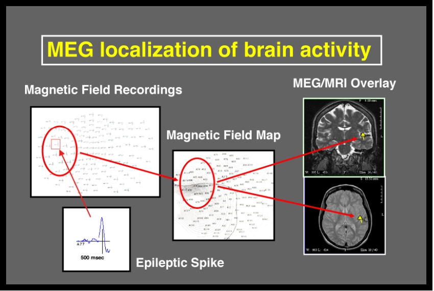 Photo source: web.mit.edu/kitmitmeg/whatis.html |
|
|
|
Post by francesco_negri on Feb 12, 2016 3:36:04 GMT -5
I'd like to know your opinion about this functional imaging techinque, called EROS (Event-Related Optical Signal).
"Event-related optical signal (EROS) is a brain-scanning technique that uses infrared light through optical fibers to measure changes in optical properties of active areas of the cerebral cortex. Whereas techniques such as diffuse optical imaging (DOI) and near-infrared spectroscopy (NIRS) measure optical absorption of haemoglobin, and thus are based on blood flow, EROS takes advantage of the scattering properties of the neurons themselves, and thus provide a much more direct measure of cellular activity. EROS can pinpoint activity in the brain within millimeters (spatially) and within milliseconds (temporally). Currently, its biggest limitation is the inability to detect activity more than a few centimeters deep, which thus limits this fast optical imaging to the cerebral cortex. EROS is a new, relatively inexpensive technique that is non-invasive to the test subject." from Wikipedia Wikipedia link. Developers site link. Paper link. Video link. Hope this can help!  |
|
|
|
Post by Vortex | Head Admin on Feb 12, 2016 16:00:55 GMT -5
I'd like to know your opinion about this functional imaging techinque, called EROS (Event-Related Optical Signal).
"Event-related optical signal (EROS) is a brain-scanning technique that uses infrared light through optical fibers to measure changes in optical properties of active areas of the cerebral cortex. Whereas techniques such as diffuse optical imaging (DOI) and near-infrared spectroscopy (NIRS) measure optical absorption of haemoglobin, and thus are based on blood flow, EROS takes advantage of the scattering properties of the neurons themselves, and thus provide a much more direct measure of cellular activity. EROS can pinpoint activity in the brain within millimeters (spatially) and within milliseconds (temporally). Currently, its biggest limitation is the inability to detect activity more than a few centimeters deep, which thus limits this fast optical imaging to the cerebral cortex. EROS is a new, relatively inexpensive technique that is non-invasive to the test subject." from Wikipedia Wikipedia link. Developers site link. Paper link. Video link. Hope this can help! Very interesting, thank you for linking. I will be sure to look into it. |
|
|
|
Post by amoses7178 on Feb 23, 2016 13:23:07 GMT -5
I'm in agreement that traditional scanning methods are not going to cut it. But, I think the creators of SAO already realized this. In the series, a major plot point was that the Nerve Gear used microwaves to somehow have a direct connection to the nerves of the brain. This was turned into a weapon of sorts that kept a virtual microwave gun pointed at the heads of 10,000 people all at once. It goes to show the brilliant writing at the very least because, although we do not yet have the science to non-invasively stimulate a nerve directly with the kind of accuracy we need to enable Full Dive VR, we DO have the ability to stimulate nerves deep within the brain in generally targeted areas using magnetic fields. A perfect example of this in real-world use is a technology called Transcranial Magnetic Stimulation ( goo.gl/sJMTfG) which uses overlapping magnetic fields to encourage targeted bundles of nerves to fire repeatedly. If a technology such as this could be miniaturized, made far more accurate, and with faster impulses, it would be perfect to both stimulate and read the deep nerves within the body while being non-invasive (ie, you don't have to install a Ghost in the Shell like port for it to work). You wouldn't have to penetrate the whole brain for it to work either. If you targeted just the cranial nerves leading into and out of the brain (which are attached to the brain stem), you could effectively "tap" our input/output to the real world, and supplant the real world with a virtual one just like they did in SAO. It would be a bit like sending your game console output through your DVR before it goes to the TV. Maybe, because microwaves are part of the electro-magnetic spectrum and are involved with magnetism, they thought to use that as the device that made SAO possible. -Alan |
|
|
|
Post by francesco_negri on Feb 23, 2016 13:47:46 GMT -5
TMS is mainly used to "write" impulses inside the brain. At the moment the best electro-magnetic reader for the brain is MEG, in fact in order to read brainwaves you need a very huge magnet and a shielded room, to prevent interferences. Actually TMS would be an interesting technique to write input signals. This is only my opinion |
|


